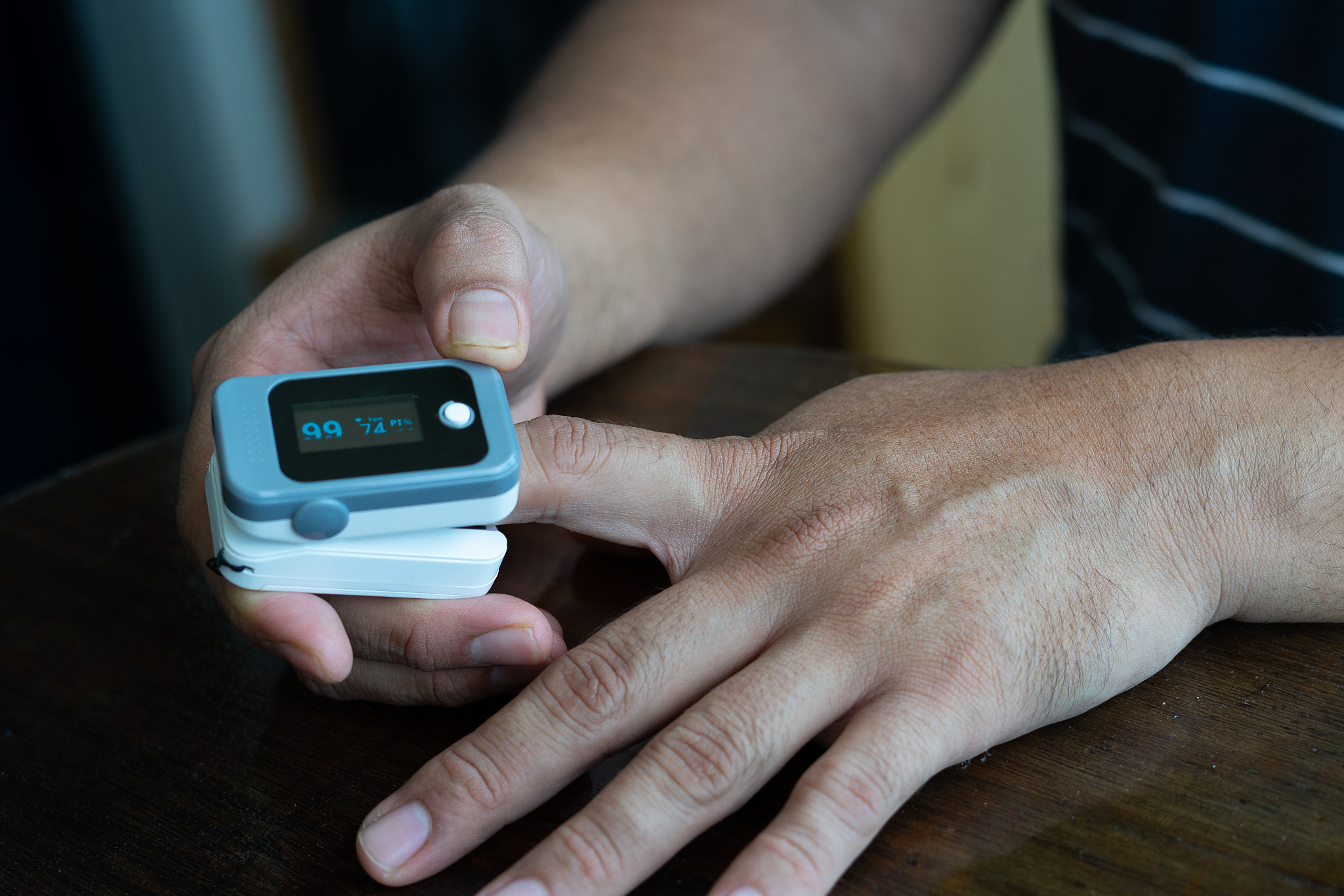
Galaxy Watch 7 May Finally Bring Blood Sugar Monitoring
페이지 정보
작성자 Robt 작성일25-09-08 17:42 조회9회 댓글0건관련링크
본문
Based on a brand new report out of South Korea, Samsung is going to introduce blood sugar monitoring with the Galaxy Watch 7 this 12 months. Hon Pak, vice president and head of digital healthcare at Samsung Electronics, highlighted the company's work on achieving noninvasive blood sugar monitoring via its wearable devices again in January this yr. He pointed out that was Samsung was placing in "significant investment" to make that happen. Pak recently met with the advisory board members of the Samsung Health platform at the Samsung Medical Center in Seoul. The discussions focused on blood sugar monitoring, diabetes, and the applying of AI to Samsung Health. The expectation now could be that Samsung will add blood sugar monitoring to the upcoming Galaxy Watch 7 collection. However, the corporate might choose to classify the smartwatch as an electronic system as a substitute of a medical gadget, largely attributable to regulatory considerations. There's additionally the chance that this function could also be made obtainable on the Samsung Galaxy Ring as effectively, the company's first good ring, that is additionally expected to be launched later this year. Whether that happens with the primary iteration product stays to be seen. It's potential that Samsung could retain some superior performance for the second iteration of its good ring. Based in Pakistan, his interests embody know-how, finance, Swiss watches and Formula 1. His tendency to put in writing lengthy posts betrays his inclination to being a man of few words. Getting the One UI 8 Watch replace? 2025 SamMobile. All rights reserved.

We efficiently demonstrated the feasibility of the proposed methodology in T2-weighted practical MRI. The proposed method is especially promising for cortical layer-specific useful MRI. For the reason that introduction of blood oxygen stage dependent (Bold) contrast (1, 2), useful MRI (fMRI) has turn out to be one of the most commonly used methodologies for neuroscience. 6-9), by which Bold effects originating from larger diameter draining veins can be considerably distant from the actual websites of neuronal activity. To simultaneously achieve high spatial resolution whereas mitigating geometric distortion inside a single acquisition, interior-volume choice approaches have been utilized (9-13). These approaches use slab selective excitation and refocusing RF pulses to excite voxels within their intersection, and restrict the sphere-of-view (FOV), wherein the required variety of phase-encoding (PE) steps are diminished at the identical decision in order that the EPI echo practice size turns into shorter along the section encoding route. Nevertheless, BloodVitals review the utility of the inner-volume primarily based SE-EPI has been limited to a flat piece of cortex with anisotropic decision for covering minimally curved gray matter space (9-11). This makes it challenging to search out purposes past primary visual areas particularly within the case of requiring isotropic high resolutions in other cortical areas.
3D gradient and spin echo imaging (GRASE) with internal-volume selection, which applies multiple refocusing RF pulses interleaved with EPI echo trains in conjunction with SE-EPI, alleviates this drawback by allowing for prolonged quantity imaging with excessive isotropic resolution (12-14). One major concern of utilizing GRASE is image blurring with a large point unfold function (PSF) in the partition course as a result of T2 filtering effect over the refocusing pulse train (15, 16). To cut back the image blurring, a variable flip angle (VFA) scheme (17, measure SPO2 accurately 18) has been incorporated into the GRASE sequence. The VFA systematically modulates the refocusing flip angles so as to sustain the signal energy throughout the echo prepare (19), thus growing the Bold sign changes in the presence of T1-T2 blended contrasts (20, 21). Despite these benefits, VFA GRASE nonetheless results in vital loss of temporal SNR (tSNR) as a result of diminished refocusing flip angles. Accelerated acquisition in GRASE is an interesting imaging possibility to scale back both refocusing pulse and EPI practice length at the identical time.
 In this context, accelerated GRASE coupled with picture reconstruction strategies holds great potential for either reducing picture blurring or bettering spatial volume alongside both partition and part encoding directions. By exploiting multi-coil redundancy in indicators, parallel imaging has been successfully utilized to all anatomy of the physique and measure SPO2 accurately works for both 2D and BloodVitals SPO2 3D acquisitions (22-25). Kemper et al (19) explored a mix of VFA GRASE with parallel imaging to extend quantity protection. However, the restricted FOV, localized by only a few receiver coils, doubtlessly causes high geometric factor (g-factor) values as a result of ill-conditioning of the inverse drawback by together with the big variety of coils which might be distant from the region of interest, thus making it difficult to realize detailed sign evaluation. 2) signal variations between the identical section encoding (PE) traces across time introduce picture distortions during reconstruction with temporal regularization. To deal with these issues, Bold activation needs to be individually evaluated for both spatial and temporal traits. A time-collection of fMRI photographs was then reconstructed under the framework of sturdy principal element analysis (okay-t RPCA) (37-40) which might resolve presumably correlated info from unknown partially correlated photos for measure SPO2 accurately reduction of serial correlations.
댓글목록
등록된 댓글이 없습니다.