Understanding Nuclear Heart Scan Data
페이지 정보
작성자 Brianna 작성일25-04-23 03:33 조회10회 댓글0건관련링크
본문
A nuclear heart scan, also known as myocardial perfusion imaging, is a diagnostic tool used to visualize the heart's function and examine coronary arteries. To understand the anatomical correlates of a nuclear heart scan, it is essential to have a basic knowledge of the heart's structure and how blood circulates.
The heart is divided into four chambers: the right and left atria, which receive blood and prepare it for the lungs, and the right and left ventricles, which pump cardiovascular fluids.
The coronary arteries supply blood to the heart muscle itself, with the left main coronary artery branching into the left anterior descending (LAD) and the left circumflex (LCx) arteries, allowing the heart to pump efficiently. The right coronary artery (RCA) supplies the right ventricle, the right atrium, and the SA node, regulating heart pace.
A nuclear heart scan involves injecting a small amount of radioactive tracer into the bloodstream. The tracer collects in the heart muscle cells, allowing a gamma camera to capture clear images. The cardiac PET (positron emission tomography) scan or اسکن هسته ای SPECT (single photon emission computed tomography) scan can create precise pictures of the heart, showing normal and abnormal areas.
When interpreting a nuclear heart scan, several key areas are analyzed. The left ventricle, which pumps blood to the body, should show uniform tracer uptake. Abnormalities in this area, such as a fixed defect, indicate scar tissue from a previous heart attack. The right ventricle, which pumps blood to the lungs, typically has reduced blood flow, and usually appears smaller.
The SA node, which regulates heartbeats and rhythm, should also show regular tracer uptake. A reduction in uptake here may indicate heart failure. In some nuclear heart scans, coronary artery locations can be pinpointed, especially for the left anterior descending or circumflex, and also the right coronary arteries based on the appearance of blood flow when comparing to a heart-blood supply anatomical schema.
The results of a nuclear heart scan provide vital data for diagnosing heart conditions. Understanding the anatomical correlates of a nuclear heart scan can help patients understand their heart.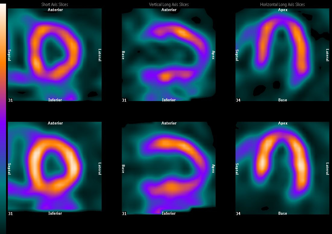 This diagnostic tool is essential for maintaining cardiovascular well-being.
The heart is divided into four chambers: the right and left atria, which receive blood and prepare it for the lungs, and the right and left ventricles, which pump cardiovascular fluids.
The coronary arteries supply blood to the heart muscle itself, with the left main coronary artery branching into the left anterior descending (LAD) and the left circumflex (LCx) arteries, allowing the heart to pump efficiently. The right coronary artery (RCA) supplies the right ventricle, the right atrium, and the SA node, regulating heart pace.
A nuclear heart scan involves injecting a small amount of radioactive tracer into the bloodstream. The tracer collects in the heart muscle cells, allowing a gamma camera to capture clear images. The cardiac PET (positron emission tomography) scan or اسکن هسته ای SPECT (single photon emission computed tomography) scan can create precise pictures of the heart, showing normal and abnormal areas.
When interpreting a nuclear heart scan, several key areas are analyzed. The left ventricle, which pumps blood to the body, should show uniform tracer uptake. Abnormalities in this area, such as a fixed defect, indicate scar tissue from a previous heart attack. The right ventricle, which pumps blood to the lungs, typically has reduced blood flow, and usually appears smaller.
The SA node, which regulates heartbeats and rhythm, should also show regular tracer uptake. A reduction in uptake here may indicate heart failure. In some nuclear heart scans, coronary artery locations can be pinpointed, especially for the left anterior descending or circumflex, and also the right coronary arteries based on the appearance of blood flow when comparing to a heart-blood supply anatomical schema.
The results of a nuclear heart scan provide vital data for diagnosing heart conditions. Understanding the anatomical correlates of a nuclear heart scan can help patients understand their heart.
This diagnostic tool is essential for maintaining cardiovascular well-being.댓글목록
등록된 댓글이 없습니다.